
Skull Fracture in an Infant Not Visible with Computed Tomography The Journal of Pediatrics
Definition. A skull x-ray is a picture of the bones surrounding the brain, including the facial bones, the nose, and the sinuses.. Alternative Names. X-ray - head; X-ray - skull; Skull radiography; Head x-ray. How the Test is Performed. You lie on the x-ray table or sit in a chair.
A Baby Xray What To Expect And How To Prepare Kidadl
Ultrasound. hip : figure 1 example normal-pediatric- hip-ultrasound-graf-type-i. Skeletal survey. Skeletal surveys are performed in cases of:. suspected non-accidental pediatric skeletal injury. 1-month-old: example 1 5-month-old: example 1 post-mortem before an autopsy in cases of suspected sudden infant death syndrome (SIDS) to exclude traumatic skeletal injury or skeletal abnormalities.

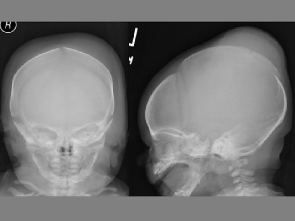
Lateral skull xray ofan 1I month old baby who allegedly fell offa... Download Scientific Diagram
The art of interpreting skull radiographs is slowly being lost as trainees in radiology see fewer plain radiographs and depend more heavily on computed tomography and magnetic resonance imaging. Nevertheless, skull radiographs still provide significant information that is helpful in finding pathologic conditions and appreciating their extents. Abnormalities in the skull may be reflected as.

Lateral skull xray in a 4 month old male. The skull is scaphocephalic... Download Scientific
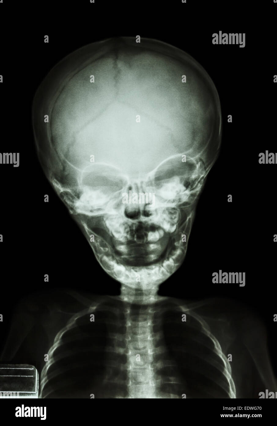
But a baby x-ray is a quick and painless way to obtain important imaging of your infant's body. While radiation exposure is a part of x-ray technology, an occasional x-ray is deemed safe for babies. This helpful tool can quickly determine the cause of sickness, injury or pain, which can outweigh any risks related to the procedure.
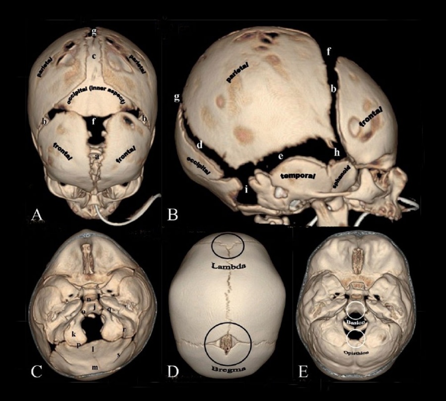
Neural Exam Newborn head shape and sutures Embryology
Introduction. Skull lesions in the paediatric population are common entities and often constitute a diagnostic dilemma for radiologists. A wide spectrum of lesions exists, which includes congenital, traumatic, infectious, neoplastic, vascular, and post-surgical abnormalities during imaging pathways.
Xray of baby’s head r/interestingasfuck
A skull X-ray is typically done after a traumatic head injury. The X-ray allows your doctor to inspect any damage from the injury. Other reasons you may undergo a skull X-ray include.

The Infant Skull A Vault of Information RadioGraphics
Bickle I, Normal AP skull radiograph - pediatric. Case study, Radiopaedia.org (Accessed on 13 Dec 2023) https://doi.org/10.53347/rID-46691

The Infant Skull A Vault of Information RadioGraphics
Sinus infection ( sinusitis) Sometimes skull x-rays are used to screen for foreign bodies that may interfere with other tests, such as an MRI scan. A CT scan of the head is usually preferred to a skull x-ray to evaluate most head injuries or brain disorders. Skull x-rays are rarely used as the main test to diagnose such conditions.
Infant skull hires stock photography and images Alamy
Indications. This examination is able to assess for medial and lateral displacements of skull fractures, in addition to neoplastic changes and Paget disease. Note: As this view results in higher radiation dose to the radiosensitive lens of the eyes compared to the PA view, it should only be used in situations where the patient is unable to face.
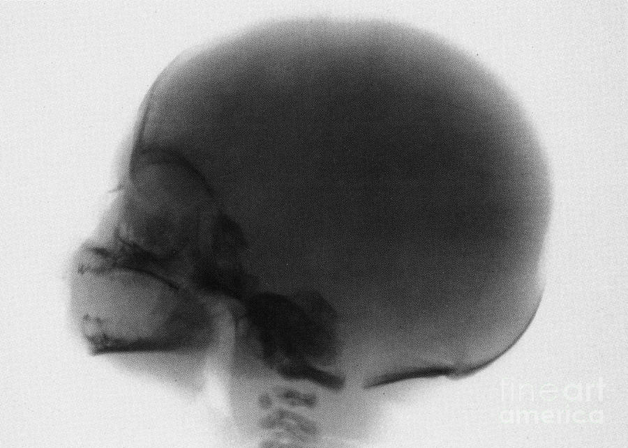
Infant Skull Xray Photograph by Photo Researchers Pixels
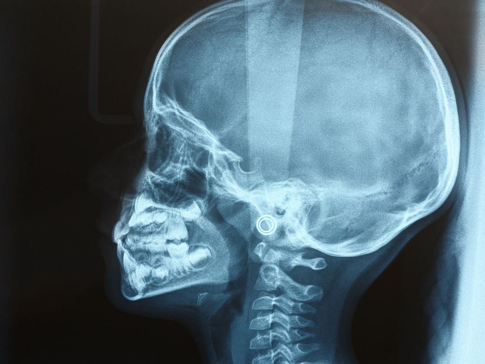
A skull X-ray is a series of pictures of the bones of the skull. Skull X-rays have largely been replaced by computed tomography (CT) scans. A skull X-ray may help find head injuries, bone fractures, or abnormal growths or changes in bone structure or size. The bones of the skull are normal in size and appearance.

Infant Skull X Ray My XXX Hot Girl
A skull X-ray works by allowing your doctor to see the bones of the skull and other tissues or foreign objects inside your head. Each part of your body absorbs different amounts of radiation.

X Ray Della Vista Laterale Del Cranio Umano Del Bambino Fotografia Stock Immagine di umano
Aim: To determine optimal exposure parameters when performing digital skull radiographs in infants with suspected non-accidental injury (NAI). Method: Anteroposterior and lateral post-mortem skull radiographs of six consecutive infants with suspected NAI were made at six exposure levels for each projection. Entrance surface doses ranged from 75-351 microGy.

The Infant Skull A Vault of Information RadioGraphics
While somewhat gnarly, sure, but it's still rather fascinating to see what it looks like as permanent teeth form within the skull before pushing out baby teeth. Below, a time-lapse video of permanent teeth growing in:
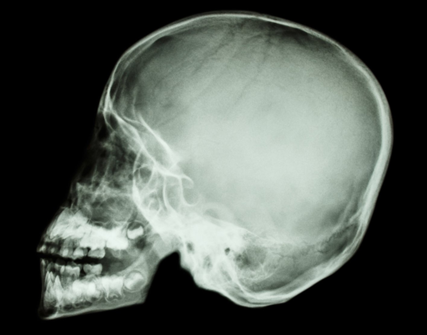
Skull xray
Newborn baby skull x-ray teeth can detect dental problems that may not be visible to the naked eye. For example, they can identify tooth decay, cavities, and other abnormalities in your baby's mouth that may require treatment. Early detection is key in preventing future oral health issues. By catching dental problems early on, you can take.

Massive congenital depression of neonate’s skull ADC Fetal & Neonatal Edition
X-rays use a small amount of radiation beams to make images. Standard X-rays are done for many reasons. They are done to diagnose tumors, infection, foreign bodies, or bone injuries. X-ray beams pass through body tissues onto treated plates. The more solid a structure is, the whiter it appears on the film.
EPOS™
X-rays use invisible electromagnetic energy beams to make images of the skull. Standard X-rays are done for many reasons, including diagnosing tumors, infection, foreign bodies, or bone injuries. X-rays use external radiation to produce images of the body, its organs, and other internal structures to diagnose a problem.